|
|
 |
Hydatiform moles or gestational throphoblastic tumors
|
 |
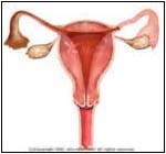
Normal |

Abnormal |
|
|
- In one out of every 1500-2000 pregnancies, a grapelike cluster of tissue forms around the area that normally would become the placenta. The placenta is where the fertilized egg attaches to the uterus (womb). The grapelike mass, called a Hydatiform mole, may grow, damage, and destroy the developing embryo (future baby).
- The majority (80%) of the moles are not aggressive, and remain in the uterus. Of the rest, 15% are aggressive and invade the wall of the uterus, causing bleeding to occur. In 2% of the cases, the moles can become cancerous and spread to other sites in the body. These cancers are called Choriocarcinoma.
|
 |
-
Vaginal spotting or bleeding.
- Nausea and vomiting.
- Rapid growth of the uterus
- Abdominal pain
- Cannot feel the fetus or baby moving.
- Invasion of the uterine wall can cause internal bleeding.
- If the mole is cancerous and spreads to the lungs, there can be coughing of blood or shortness of breath and pain.
- Some moles can produce thyroid like hormones that bring about symptoms of thyroid disease.
- There can be Preeclampsia, which occurs in some normal pregnancies (after 20 weeks), consisting of High Blood Pressure, fluid retention, and loss of protein in the urine. In Molar Pregnancy, Preeclampsia occurs under 20 weeks of pregnancy.
|
 |
- The Hydatiform mole forms from a small piece of the placenta left from a previous pregnancy or Miscarriage.
|
 |
- The physical exam includes a Pelvic exam (Examination of the vagina and the uterus by the doctor) that may show a larger than expected uterus.
- There may be High Blood Pressure.
- There is an elevation of the pregnancy hormone Beta HCG (Human Chorionic Growth Hormone) in the blood.
- A Sonogram or Ultra sound uses sound waves to show the mole as a cluster of grapes or as a stormy appearance.
- The Sonogram may also show absent
fetal heart movement and heart sound.
|
 |
- Previous Miscarriage
- Normal pregnancy
- Ectopic (outside the uterus) pregnancy
- Age over 40
- Geographic location -- Women from Mexico, Southeast Asia, and the Philippines are at higher risk than White American women
|
 |
-
Best
treatment for benign moles. This removes them with
dilation, evacuation, or Curettage (D&E and D&C), in
which a vacuum device is used to suction the mole
out.
- To assure that the mole is gone and did not return after the D&E, the following is done:
- The mole is examined by a specialist (pathologist) under the microscope to rule out cancer.
- A weekly measurement of the BHCG is preformed until it reaches a value greater than 100mIU/ml.
- BHCG every 2 weeks for 3 months and then monthly for one year
- There is a Pelvic exam every 2 weeks until the uterus size returns to normal, then every 3 months for the following year.
- A chest X-Ray should be done every 4-6 weeks to make sure that there is not a cancerous spread to the lungs.
- If there is elevation of BHCG two weeks after the mole is removed, this may indicate the presence of Choriocarcinoma (cancerous), or a spread of the cancerous mole and further evaluation using an MRI (pictures of body taken using magnetic energy), Cat Scan, or a Sonograph.
- The treatment of an invasive and a cancerous mole may include a hysterectomy (removing the uterus) and the use of Chemotherapy (cancer killing medications).
- Sometimes, Radiation energy is used if the Choriocarcinoma has spread to the brain.
- The patient is always put on birth control, and must not get pregnant for one year.
|
 |
-
Work with your physician and do not loose hope. If the diagnosis is made early, the chance for future pregnancies (even if the mole is cancerous or invasive) is good.
|
| | |
If you want your friend to read or know about this article, Click here
 |
|
|