|
|
 |
Retinal vein occlusion
|
 |
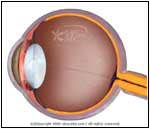
Normal |
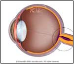
Abnormal |
| |
- Retinal Vein Thrombosis refers to blockage of the central retinal vein that carries blood away from the eye. Occlusion of this vein or its branches can be seen in elderly, especially in those with Glaucoma, Diabetes Mellitus, and Hypertension.
- Occlusion due to a clot (thrombosis) may be seen in those with increased risk of thrombosis (e.g., sickle cell disease), increased blood viscosity, and an elevated blood Hct (hematocrit).
- Painless visual loss is often the only symptom. Upon examination, the retinal veins appear distended and tortuous, the fundus of the eye appears congested and swollen, and numerous hemorrhagic areas may be seen on the retina in the back of the eye. These changes may be limited to one quadrant, if the obstruction involves only a branch of the vein. New vessels (neo-vascularization) may form in the retina and secondary (neovascular) Glaucoma can occur weeks after the occlusion.
- Tests such as fluorescein Angiography may help in assessing the circulation of the eye and the status of the blockage.
- Patients with thrombosis may be at risk for developing clots in other parts of the body (legs, brain, heart) and may need further evaluation by an ophthalmologist and their regular physician.
- Procedures (such as photocoagulation to remove new vessels formed) can prevent secondary neovascular Glaucoma.
|
| | |
If you want your friend to read or know about this article, Click here
 |
|
|